In 2023, I suddenly went blind. That was a fun day. Not many things cause that, none good.
It could have been worse.
Turns out, my body tried to eat my optic nerves, but I won. 20/20 vision baby, and slim to zero chance of recurrence. I have a rare autoimmune disorder, MOGAD, and some of the worlds best studying/fighting it are right here in Australia. I can't thank about 8 seperate teams at the Royal Melbourne hospital enough.
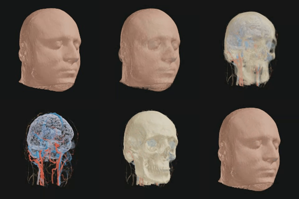
Years later, one day I got bored. "Wait, I have a dvd with a scan of my brain, and I play with data". Could I interpolate something and build a custom model? 15,191 DICOM MRI images later...
The ultimate goal? Print a 3d model. That flashes for various reasons. Maybe when there's lightning. Or I get a quad-kill in Wardogs. Or when my wife's business gets another big, tasty order (that's happening a lot).
The thought of getting stimulus from my brain but smol, tickles me. There's something about it being visual stimulus, no less, thats deeply, darkly ironic. (Fuck you, MOGAD. I win.)
once we have it, we can do some iiinterestin things. Animations. More interactivity. Maybe an educational visualisation of what MOGAD was up to. Etc.
About the project
From scan slices to a portrait.
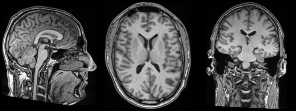
This portrait uses the head study, acquired on a Siemens MAGNETOM Sola 1.5 T MRI scanner at the Royal Melbourne hospital
A lot of those 15,191 images show the same anatomy at different times or from different angles. They aren’t all separate scans with new detail. For example, the angiography sequence contains 40 original volumes and 39 background-subtracted volumes following contrast through the vessels.
The images we used
- T1 MPRAGE, before and after contrast: two 208-slice volumes provide the main head and brain reference, with approximately 1 mm stored spacing.
- TWIST contrast angiography: the first nine subtracted phases provide the main vessel signal. All 39 subtracted phases were reviewed; their timing helps estimate arterial and venous groups.
- T2, FLAIR and susceptibility-weighted images: complementary views help cross-check brain, fluid and vessel boundaries.
- Targeted eye scans, including STIR and Dixon-water: supporting views help estimate the eye outlines. Eventually this will become a higher fidelity focus area.
How it comes together
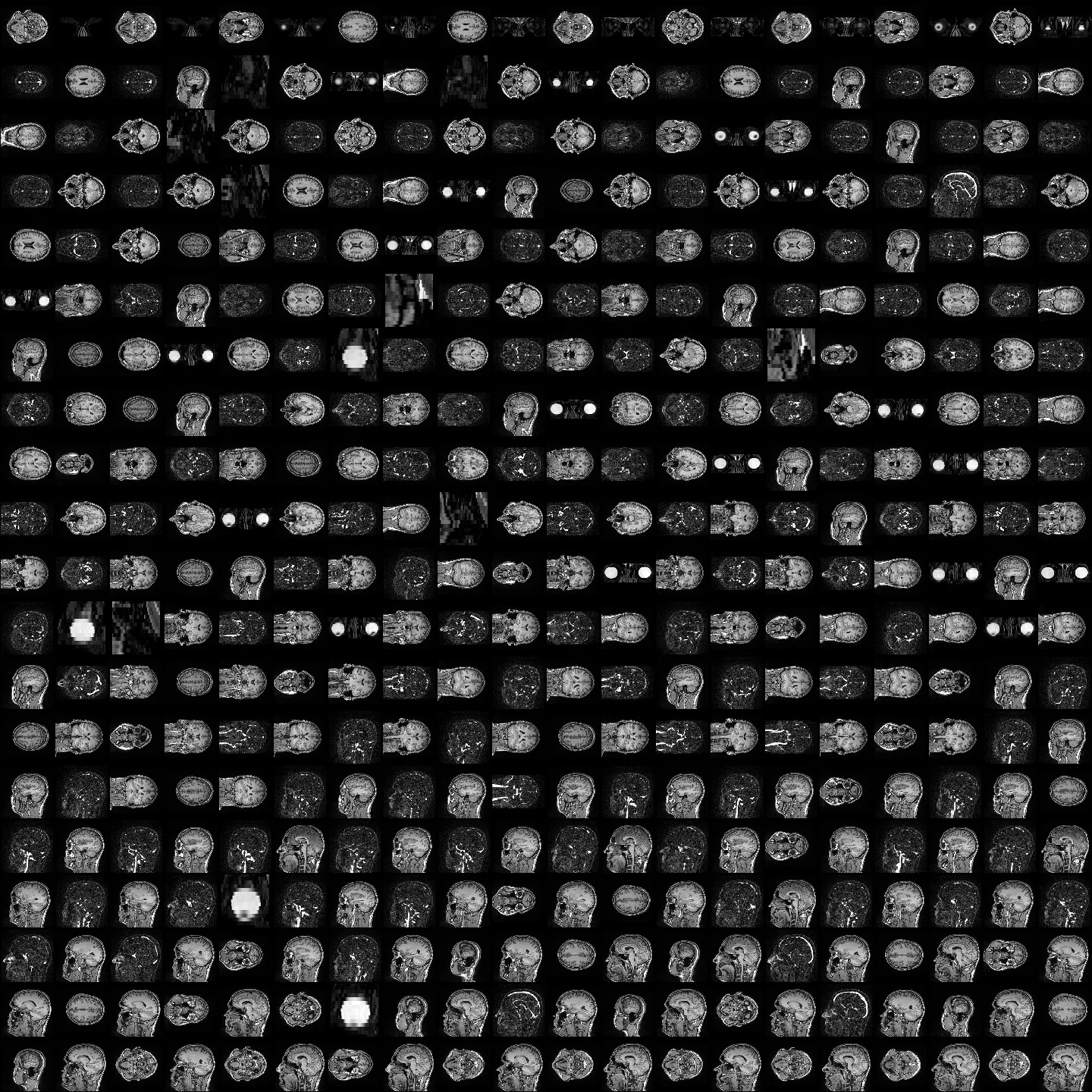
Slices were ordered by their recorded position and assembled into 3D volumes. Compatible head scans were re-aligned in the same physical space, and compared to estimate the tissue layers. Vessel signal is combined across the early contrast phases; different MRI contrasts support different structures, rather than being blindly added together.
The resulting volumes are compressed for the browser. The viewer samples through them to build the image you see, with separate colour and opacity controls. FreeSurfer helps estimate brain tissues; these boundaries and the vessel labels remain interpretations of the scans.
What’s being worked on
A more performant rendering method.
More refined detail from integrating more studies/scans.
Smoothing between slices.
A.I generated best-guest fill in where gaps exist (already lightly leveraged).
A part / structure picker and index, highlighting system.
More performant rendering for mobile devices.
Finer vessel continuity and tissue boundaries.
Then the fun stuff.
Music-synced pulsing through veins/nerves/structures, affecting transparency, etc.
Material swapping. Turn the brain to glass. Veins to sand. Etc.
Whatever you come up with.
Credits + software used
SUPPORT THE THINGS I MAKE
Donate
I love making things. Anything I've ever built has been to have fun, share fun, and make life a bit easier. If you got value from one of these things, and you'd like to chuck us a coffee, a bottle, or a god damned Ferrari, go your hardest. Then hustle over to discord to claim your supporter role!
- GitHub Sponsors
- Buy Me a Coffeeguest checkout available
- Donate with PayPal
More things I made: browse the portfolio